FFR: Other Experiments
Supporting original reading. Follow the AP topic pathway for reviewed scope, worked reasoning and targeted practice.
Chemistry II · original Chapter 4 · combined Chapter 12
Section Breakdown
- Synthesis of Aspirin
- Beer’s Law
- Testing the Purity of Aspirin
Section 4
FFR: Other Experiments
Interactive 4.4.1 Aspirin
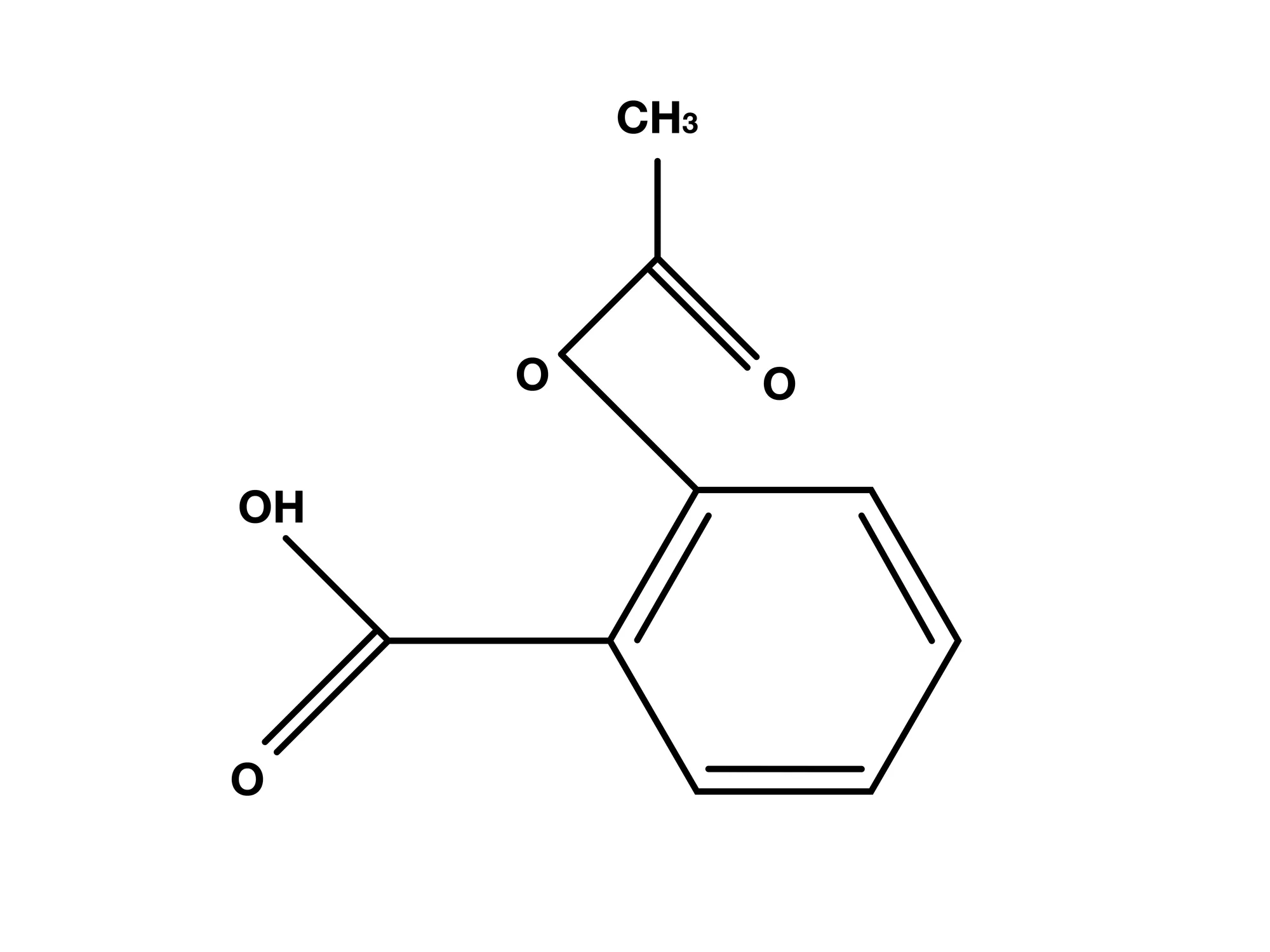
Read all original figure notes
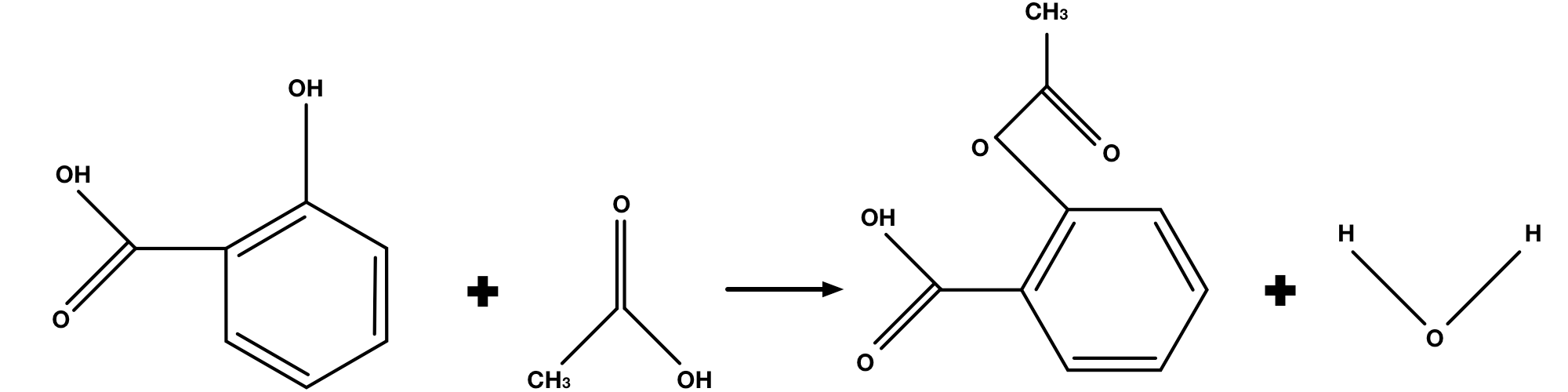
Organic synthesis enrichment: aspirin is acetylsalicylic acid. Its aromatic ring bears a carboxylic-acid group and a phenolic-oxygen acetyl ester, −O−C(=O)−CH₃. A methyl group is part of the acetyl group, but this is not a methyl ester of the carboxylic acid. Follow the bonds when naming the functional group.
Aspirin can be synthesized from salicylic acid and acetic acid. This process uses the following equation:
It is easier to identify what is going on if you look at the lewis diagram version of the reaction.
Salicylic acid has a phenolic hydroxyl group, −OH, rather than a free hydroxide ion, OH⁻. Acetylation replaces that hydroxyl’s hydrogen with an acetyl group while retaining the oxygen in the ester linkage. The aspirin product contains a phenolic-oxygen acetate ester, not a methyl ester. These functional-group details are enrichment beyond the required AP targets.
Notice that the reaction between salicylic acid and acetic acid produces water. This is not very desirable, as this means that if we are to perform this reaction in water (which is preferable, as it encourages the molecules to interact), the water that is present can react with aspirin to push the equation to the left, producing salicylic acid again.
Acetic anhydride is an acid anhydride, CH₃C(=O)−O−C(=O)CH₃, not an ester made of two methyl groups. In the standard conceptual aspirin synthesis, salicylic acid receives an acetyl group and acetic acid is a coproduct. An actual experiment uses an instructor-reviewed protocol specifying reagent amounts, temperature control, ventilation and waste handling.
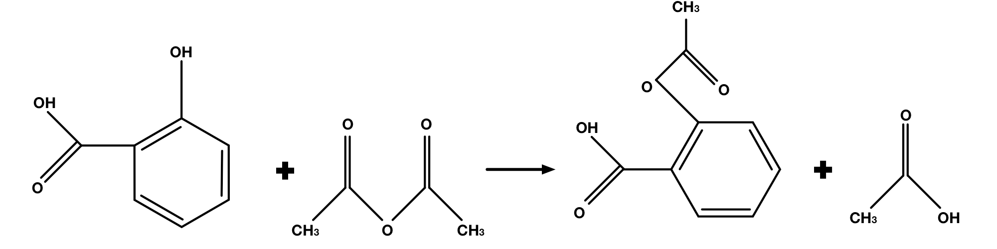
Producing acetic acid rather than water does not make aspirin immune to hydrolysis. Aspirin’s ester can react with water to produce salicylic acid and acetic acid, and the rate depends on conditions including temperature and pH. Excess acetic anhydride also reacts with water. Keep product chemistry and the approved isolation/quench procedure separate.
So how do we go about actually synthesizing the Aspirin? The lab procedure can vary depending on what your instructor decides to concentrate on, so the lab outline we provide will try to cover as many aspects of Aspirin synthesis as we can.
In order to synthesize Aspirin, most labs will use the reaction we show above, that is, Acetic Anhydride and Salicylic Acid. Now, this reaction is not the fastest, so we will use Sulfuric Acid and heat to speed it up. Sulfuric Acid acts as a catalyst for the Aspiring synthesis, and as we have shown before, heat tends to speed up reactions in aqueous systems.
The actual numbers will vary depending on how much Aspirin you want to create, so you will have to rely on your instructor to tell you how much of each component you need.
You will begin with setting up your experiment. This will probably be the bulk of your time spent on this lab, as the synthesis itself gets pretty quick by adding the heat and the sulfuric acid.
Before you can begin adding any chemicals, you need to make sure you have all of your necessary equipment and reagents.
- (2) Beakers large enough for a water bath
- (1) Erlenmeyer Flask
- (1) Bunsen Burner with Beaker Stand
- (2) small Graduated Cylinders (can get away with 1 if needed)
- Buchner Funnel or other filtration set-up
- Scale
- Hot Plate
- Access to Fume Hood
- Supply of Deionized Water, perhaps a spray bottle for it
- Acetic Anhydride (liquid form)
- Salicylic Acid (solid form)
- Catalyst
Once you ensure you have everything you need, you need to start setting up your experiment. Throughout the course of your experiment you will need a hot water bath and a cold water bath. The cold water bath can be prepared later on, but you want to start with prepping your hot water bath. Take one of your large beakers and fill it with enough water to submerge your Erlenmeyer Flask. Then place that on a hot plate to warm. Try not to let the water boil. Keep this bath underneath the fume hood.
While that’s warming up, you can begin your experiment.
- Start by measuring out your Salicylic Acid. Make sure to note what the exact weight you use is. Add this powder to the Erlenmeyer Flask.
- Measure out your catalyst and set it aside.
- Using the graduated cylinder, measure a small amount of Acetic Anhydride and bring it and the Erlenmeyer Flask to the fume hood.
- While underneath the fume hood, add the Acetic Anhydride to the Erlenmeyer Flask containing your Salicylic Acid. Make sure to get all of the Salicylic acid into the solution, you don’t want any crystals left behind.
- Now you add in your catalyst, be it acid or sodium acetate. Once you have ensured that it entered the solution, again making sure no crystals are stranded on the sides, place the flask in your hot water bath.
- You’re going to have to wait for about 20minutes or so while the two reagents react. While that happens, use a graduated cylinder to measure out a few mLs of DI water. If you only have one graduated cylinder, ensure you clean it before measuring your DI water.
- The archived heated-anhydride water-addition instruction is superseded as a student procedure. Reaction with excess acetic anhydride can release heat, so the amount, addition rate, temperature, protective measures and ventilation must come from the instructor’s separately reviewed protocol. Do not infer a safe operating scale or quench from this draft. For the learning task, explain that hydrolysis of excess anhydride forms acetic acid and that cooling/crystallization conditions affect isolation.
- The archived heated-anhydride water-addition instruction is superseded as a student procedure. Reaction with excess acetic anhydride can release heat, so the amount, addition rate, temperature, protective measures and ventilation must come from the instructor’s separately reviewed protocol. Do not infer a safe operating scale or quench from this draft. For the learning task, explain that hydrolysis of excess anhydride forms acetic acid and that cooling/crystallization conditions affect isolation.
- While the Acetic Acid is forming, prepare your ice bath. Take the other beaker you had prepared and fill it with ice cubes and water.
- Once the reaction completes, add an excess (at least 25mL) of DI water to the solution, and then place it into the ice bath.
- Let the solution cool for about 15-20 minutes. During this time, you should start to see crystals develop as the Aspirin gets less soluble in the cold water. While you’re waiting, start cooling another 30mL or so of DI water.
- Once the 15-20 minutes has elapsed, it is time to filter your crystals.
- Set up the Buchner funnel, and pour your solution into the funnel. Make sure to get as much of the crystals as you can into the filter.
- Take the 30mL of cold DI water, and use that to wash your crystals a few times. This helps to increase the purity of your crystals.
- When your sample is washed, leave it on the Buchner funnel to air-dry for a few minutes.
- If your lab contains a method of further drying your crystals, perhaps with an oven or heat lamp, do so. This removes further moisture from your crystals, ensuring that you weigh the crystals themselves, and your measurements are not distorted by the presence of water.
- You now have Aspirin Crystals. Weigh them to see how much Aspirin you have created.
Part 2: Testing the Purity
In some labs, you will go beyond the synthesis of aspirin and test the purity of the synthesized aspirin. Throughout this lab, you synthesized acetylsalicylic acid. You may also have some solid salicylic acid crystalized amongst your Aspirin. In order to figure out how much of your crystals are Aspirin, you have quite a few options.
Two of the most common are looking at the melting point of your crystals and using spectrometry.
The primary contaminant that might be present in your sample is Salicylic Acid. Therefore you need to distinguish between the presence of Aspirin and Salicylic Acid. Luckily, the two compounds have fairly different melting points. Aspirin has the lower melting point, at 136ºC, while Salicylic Acid melts at 159ºC. Therefore, in order to check the purity of your sample, you need to identify the melting point of your crystals.
A common impurity effect is a depressed melting point and a broader melting range compared with a suitable pure reference, though mixtures and experimental conditions can behave differently. An unexpectedly high value can reflect thermometer calibration, heating rate, decomposition or another substance; it is not a general signature that impurities always raise the melting point. Use a measured range and other evidence to assess the product.
The other option you have to determine the purity is through absorbance spectroscopy. This technique provides a much more quantitative analysis of the sample’s purity.
Interactive 4.4.2 Absorbance Spectrum of the Tetraaquosalicylatroiron complex.

Read all original figure notes
How can you use light to determine the concentration of a compound, though?
Well, let’s introduce a concept called Beer’s Law. Beer’s Law, also known as the Beer-Lambert Law, quantifies the absorbance of light with respect to concentration. The law is:
There, is the measured absorbance, is the molar absorptivity, is the path length of the light, and is the molar concentration of the absorbing compound.
The molar absorptivity is unique to a compound and a wavelength. This means that the molar absorptivity of Tetraaquosalicylatroiron at 460nm is different than its molar absorptivity at 350nm.
In order to get the best measurements, we want to make sure that we have the highest response for the Tetraaquosalicylatroiron complex. This means we need to take a look at the absorbance spectrum of the compound:
As you can see, the highest absorbance is in the range of green light, 520-570nm. This means that we should set our spectrophotometer to somewhere in that range.
The spectrophotometer will pass light at a given wavelength through our sample, and detect how much of the light is absorbed.
If you want to know how the Absorbance is calculated, it is based off the intensity of the light before and after it passes through the sample. For liquids, this is calculated by:
Absorbance is A=log₁₀(I₀/I)=−log₁₀(I/I₀), where I₀ is reference incident intensity and I is transmitted intensity. Transmittance is T=I/I₀. Therefore a sample transmitting less light has larger positive absorbance. The reciprocal inside the logarithm matters; log₁₀(I/I₀) would give the wrong sign.
Where is the absorbance, is the final intensity, and is the initial intensity. Using this relationship, the spectrophotometer reads out to us the absorbance of our sample.
Now we can measure the absorbance of our sample after we add some iron, right?
Strictly speaking, yes, you can. However, without the molar absorptivity, this one number won’t do you much good. The molar absorptivity will change with every compound and every wavelength, meaning that there are no tables for you to just look up the molar absorptivity of a compound at a wavelength. Absorbance can also change depending on other compounds in solution, meaning that you can have a situation which results in higher absorbances than you expect due to two compounds absorbing the same light. Instead, we need to determine the molar absorptivity ourselves.
In order to determine an unknown absorptivity, we need to know everything else in Beer’s Law. This means knowing the Absorbance, the path length, and the Concentration. The path length will be constant throughout your measurements, so it might be easier to couple that with your molar absorptivity, and just call the equation , where represents the impact of molar absorptivity and path length together, as both of these will be constant in our experiment. We will call this the coefficient of absorbance.
So how can you figure out the concentration of an unknown sample?
The easiest way is to compare it to known samples. Beer’s Law tells us that we should have a linear relationship between concentration and absorbance. Therefore, if you draw up a relationship by measuring known concentrations, you can determine the coefficient of absorbance for your set-up.
Reviewed supplied-data calibration exercise: an unspecified dilute colored solute is measured at a selected wavelength with matched1.00cm cells. All standards and the unknown have the same solvent/reagent matrix and measurement procedure. This replaces the unsupported copper-chloride wavelength and very concentrated stock. We will interpret the supplied data rather than certify an unreviewed chemical recipe.
For the supplied model, use a100µmolL⁻¹ stock to prepare standards with final volumes10.00mL. Stock aliquots0,1.00,2.00,3.00and4.00mL give concentrations0,10.0,20.0,30.0and40.0µmolL⁻¹ after final-volume dilution. Use a matrix-matched blank and instructor-approved calibrated technique. The original1–10M series is superseded; linearity must be evaluated within the stated measurement range.
The following are newly supplied model observations, not measurements recovered from the source. Concentrations0,10,20,30,40µmolL⁻¹ give absorbances0.008,0.258,0.508,0.758,1.008. The unknown has absorbance0.633. Plot the standards, fit slope and intercept, and check whether the unknown lies inside the calibration range.
| Concentration/µmolL⁻¹ | Absorbance |
|---|---|
| 0 | 0.008 |
| 10 | 0.258 |
| 20 | 0.508 |
| 30 | 0.758 |
| 40 | 1.008 |
| Unknown | 0.633 |
The reviewed plot has concentration inµmolL⁻¹ on the horizontal axis and absorbance on the vertical axis. Its five points fall onA=0.0250c+0.008. The original raster graph is superseded; use the reviewed calibration plot linked here.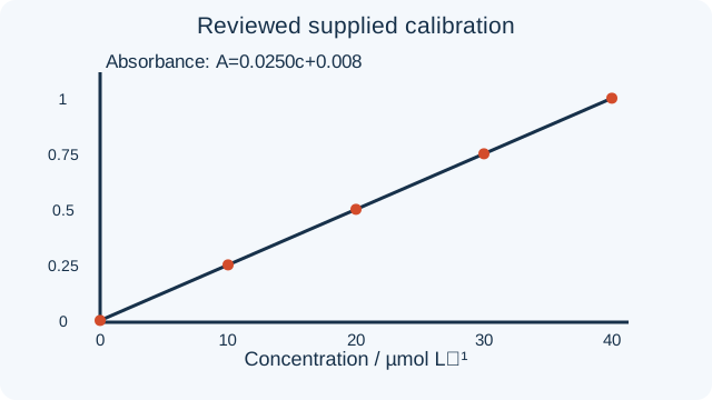
Our equation for this best-fit is:
The supplied best-fit relation is A=0.0250c+0.008, where c is inµmolL⁻¹, A is dimensionless and the slope units areLµmol⁻¹.
Do not automatically force the fitted intercept to zero. A proper blank should reduce background, but residual offsets and experimental uncertainty can remain. Examine the free-intercept fit, blank and residuals; constrain an intercept only with a justified model. Here the supplied model has intercept0.008, which remains in the unknown calculation.
Use the reviewed free-intercept calibration, A=0.0250c+0.008. The old zero-intercept comparison plot is superseded because forcing it changes the slope and can bias the inferred concentration.
A=0.0250c+0.008, with c inµmolL⁻¹. This is the reviewed supplied-data fit; it replacesA=0.0775c.
We can now use this equation to solve for our unknown sample’s concentration:
Rearrange the reviewed fit A=0.0250c+0.008 to solve for c.
c=(A−0.008)/0.0250, with c inµmolL⁻¹. Subtract the fitted intercept before dividing by slope.
For the supplied unknown, c=(0.633−0.008)/0.0250=25.0µmolL⁻¹.
The reviewed unknown concentration is25.0µmolL⁻¹, or2.50×10⁻⁵molL⁻¹. It lies inside the0–40µmolL⁻¹ standards range, so this is interpolation under the stated model.
We prepared a stated dilute calibration, used matched measurement conditions, fit slope and intercept, then calculated the unknown as(A−b)/m. That is the chain of evidence. Applying a linear relation outside a validated range or forcing a zero intercept without justification can produce a precise-looking but biased concentration.
Determining Purity of Aspirin:
A color response can support a salicylate analysis under a validated method; it does not automatically determine aspirin purity. Process samples and salicylic-acid standards through the same approved procedure and verify that aspirin hydrolysis or another species does not generate the measured response.
- Sample preparation must come from the instructor’s reviewed analytical method. Choice of solvent, temperature and time affects dissolution and possible aspirin hydrolysis. For this lesson, interpret supplied processed-sample data instead of improvising a cold-water, hot-water or acetone preparation.
- When interpreting a processed sample, record the weighed sample mass, final solution volumes and each aliquot/dilution. Those quantities support a mass balance only if recovery and analyte stability have been validated under the procedure.
- Heating an aspirin-containing solution can hydrolyze aspirin and create salicylic acid during sample preparation. A method claiming to measure pre-existing salicylic-acid impurity must validate recovery and selectivity under controlled time, temperature andpH. The archived open-ended heating instruction is superseded; use an instructor-reviewed analytical procedure and appropriate controls.
- Iron(III)-salicylate color depends on solution conditions and can involve different complexes. Do not assume the source’s fixed grams-of-iron-per-gram-of-salicylic-acid ratio independently of reagent identity,pH,speciation and validated method. Calibrate against salicylic-acid standards processed through the same reviewed procedure; the absorbance measurement then measures the calibrated response under those conditions.
- The reviewed method must establish iron reagent excess, pH, matrix composition and calibrated final volume for samples and standards. Do not fill an arbitrary beaker and infer the final concentration. A stronger color is a measured response to investigate, not by itself a unique aspirin-purity result.
- Now you need to create a comparison solution.
- Use supplied salicylic-acid standards processed under the same validated conditions as the sample. Label the stock and record its analytical concentration after all preparation and reagent-addition dilutions. Do not assume that an unspecified few grams or a very dark solution establishes a linear calibration range.
- Now you need to calculate the molarity, using the following procedure:
- Begin with the mass of Salicylic Acid you got, call it .
- Determine the initial solution you created, before adding the iron. Do this by using the final volume of the solution you made and the molar mass of Salicylic Acid (138.13).
- Now determine the concentration of the salicylic acid once you dilute it with the iron solution. Recall that:
- The complex stoichiometry and quantitative formation must be established for the method’s conditions before identifying complex concentration with salicylic-acid concentration. An empirical calibration using identically processed salicylic-acid standards can avoid assuming a universal1:1complex. ControlpH,reagent excess,matrix and preparation-induced hydrolysis, and report what the method actually measures.
- Now that you know your initial concentration for the Stock Solution, you can dilute it a few times to provide a solid Beer’s Law plot.
- A stated twofold dilution can be made by transferring5.00mL to a calibrated final10.00mL volume, using the method’s matched non-analyte matrix. The resulting analytical concentration is half the original if no analyte is lost or generated. Plain water is appropriate only when the reviewed procedure establishes that the reagent conditions and response remain matched.
- Do the same procedure 3 more times, so that you have the following sets of solutions:
- Aspirin Solution
- Stock Solution A (at molarity )
- Solution B (at molarity )
- Solution C (at molarity )
- Solution D (at molarity )
- Solution E (at molarity )
- Finally, you need a blank. Perform steps 1-5 without any solids. That means, if you used acetone to dissolve the acid previously, simply start with pure acetone. If you had hot water instead, use hot water. Make sure to replicate the procedure as you had done it previously.
- This plan has five calibration levelsA–E,a blank and one unknown: six known preparations including the blank, plus one unknown. Keep concentrations and all dilution factors in a table so that the measured unknown is not accidentally counted as a known standard.
- Measure all standards, blank and unknown at the same method-validated wavelength, path length and instrument settings. The archived540nm setting is not universal for every solvent, pH or iron complex. Use the instructor’s validated measurement procedure and record the selected wavelength.
- In most cases, absorbance is measured by taking a solution and placing into a cuvet, which is a small square-ended prism.
- Many cuvets have two rough sides and two clear sides. If yours are like this, make sure to handle the cuvets by the rough sides, and do not touch the clear sides.
- Fill the cuvet with your solution that you are examining. Some machines require a blank to compare against. This will be the final solution you created, the one without any crystals added.
- Set the spectrophotometer to the selected validated wavelength, use its proper blank/reference procedure, then measure and record absorbance. Absorbance is the result, not the wavelength setting.
- Once you have the absorbance for all the solutions you created, draw up a Beer’s Law plot for the data.
- Fit absorbance against the known calibration concentrations with a freely fitted intercept first. Inspect residuals and the blank. Do not impose zero solely because a blank was used; retain an offset when supported by the measurement model and data.
- Use both the fitted slope m and intercept b inA=mc+b. Check units, calibration range and uncertainty, and include all sample-preparation dilution factors when converting back to the original sample.
- Calculate c=(Aunknown−b)/m, rather than A/m unless b=0is independently justified. This is the concentration represented by the calibration under its validated conditions, not automatically a unique chemical identity.
- Report the calibrated salicylic-acid response and its limitations. Proceed to an impurity mass fraction only if the procedure selectively measures pre-existing salicylic acid and its recovery is validated; further evidence is needed for an aspirin-purity claim.
- Call the concentration inferred from the salicylic-acid calibration cassayed acid. It is not automatically an aspirin concentration. State what the calibration and sample-preparation controls establish about the measured analyte.
- Apply each known aliquot and final-volume dilution factor to convert the assayed solution concentration back to the original prepared sample. The conservation relation c₁V₁=c₂V₂ requires no analyte loss, reaction or formation during that dilution; validate those assumptions.
- Under a selective validated salicylic-acid method, mass=moles×138.13g mol⁻¹. Include all final-volume and dilution factors to find the analyte amount represented by the original weighed sample. Heating-generated salicylic acid must not be counted as pre-existing impurity without correcting for it.
- If a selective validated method gives the mass of pre-existing salicylic-acid impurity, report100msalicylic acid/msample. Subtracting that percentage from100does not by itself prove the remainder is aspirin: other impurities and preparation-induced hydrolysis must be excluded by additional evidence.
- You have completed a calibration-based analysis under stated assumptions. Report the measured response, dilution factors, selectivity controls and limitations. Claim aspirin purity only when the method and complementary evidence support that specific composition; a colored response alone is insufficient.
Glossary and deeper reading · 24 refreshers
These editorial refreshers supplement the reading. Definitions and examples are added guidance; original source wording and recorded answers remain in their original context.
-
Beer–Lambert law
The Beer–Lambert law relates absorbance to concentration, light-path length, and molar absorptivity under suitable conditions. Comparisons require a controlled wavelength, consistent path length, and an appropriate calibration. For example, doubling concentration doubles absorbance in the law's linear range if the other factors remain fixed.
-
Aromaticity
Aromaticity describes a special stability associated with particular cyclic, conjugated electron arrangements. Having a ring alone does not make a compound aromatic. For example, benzene is aromatic while cyclohexane is not.
-
Ester
A carboxylic ester has a carbonyl carbon bonded to an oxygen that also connects to a carbon-containing group. It differs from a carboxylic acid because that linking oxygen does not carry the acid's proton. For example, ethyl acetate is an ester containing an ethyl group attached through oxygen.
-
Lewis structure
A Lewis structure represents valence electrons using bonds and lone pairs. Its construction requires an electron count, an atom arrangement, and a final check against the total available electrons and charge. For example, water's Lewis structure has two O–H bonds and two lone pairs on oxygen.
-
Ion
An ion is an atom or group of atoms with a net electric charge. Ordinary ion formation changes electron count without changing the element's proton count. For example, a sodium atom losing one electron becomes a sodium ion with charge +1.
-
pH
pH is the negative base-ten logarithm of hydronium activity. In suitable dilute models, activity is approximated by molar concentration relative to the standard concentration. For example, lowering hydronium concentration by a factor of ten raises pH by one unit.
-
Catalyst
A catalyst participates in a reaction pathway and is regenerated overall. It can increase the reaction rate by providing a different pathway without changing the reaction's equilibrium constant at fixed temperature. For example, a catalyst can help a mixture reach equilibrium sooner without changing the final equilibrium composition.
-
Aqueous solution
An aqueous solution is a solution in which water is the solvent. The dissolved substance may originate as a solid, liquid, or gas, so aqueous does not mean that a solid has melted. For example, sodium chloride dissolved in water is aqueous even though the pure salt is solid.
-
Filtration
Filtration separates suspended particles from a fluid using a barrier that retains appropriate particle sizes. It generally does not remove individually dissolved ions or molecules. For example, a filter can separate an undissolved precipitate from the surrounding solution.
-
Deionized water
Deionized water has had most dissolved ions removed. It is not necessarily free of every nonionic impurity or microorganism. For example, using deionized water can reduce unintended ions in a prepared analytical solution.
-
Solubility
Solubility describes how much of a substance can dissolve in a specified solvent under stated conditions. It depends on the solute, solvent, temperature, and other relevant conditions and is distinct from how fast dissolution occurs. For example, two salts can have different equilibrium solubilities in water at the same temperature.
-
Mixture
A mixture contains two or more substances whose proportions can vary. Its components retain their chemical identities until a chemical reaction changes them. For example, water and ethanol can form mixtures with many different compositions.
-
Absorbance
Absorbance is a logarithmic measure of how much a sample reduces transmitted light at a specified wavelength. It depends on the ratio of incident to transmitted intensity rather than simply being the fraction absorbed. For example, absorbance 1 corresponds to transmission of one-tenth of the incident intensity.
-
Spectroscopy
Spectroscopy studies interactions between matter and electromagnetic radiation. The absorbed or emitted frequencies can reveal particular energy changes in a species. For example, infrared absorption often corresponds to changes in molecular vibration.
-
Molar absorptivity
Molar absorptivity describes a species' absorption strength at a specified wavelength in the Beer–Lambert model. It depends on the absorbing species and measurement conditions, while concentration and path length are separate factors. For example, selecting a wavelength near an absorption maximum can give a larger absorbance for the same concentration and path length.
-
Molarity
Molarity is the amount of a solute in moles divided by the final solution volume in liters. It describes concentration rather than the total amount of solute. For example, 0.10 mole dissolved to make 0.50 liter of solution gives a molarity of 0.20 mole per liter.
-
Dilution
Dilution lowers solute concentration by adding solvent while conserving the solute amount when no reaction or loss occurs. Concentration calculations use the final solution volume rather than simply the volume of solvent added. For example, increasing a solution's volume from 100 to 200 milliliters halves its concentration if its solute amount is unchanged.
-
Solute
A solute is a component dispersed in a solvent to form a solution. It need not be a solid, and a solution need not be liquid. For example, carbon dioxide is a gaseous solute in carbonated water.
-
Solvent
A solvent is the component used as the dissolving medium in a solution. It is often present in the greater amount, though the designation also depends on the mixture's context. For example, water is the solvent in a dilute aqueous sugar solution.
-
Aliquot
An aliquot is a measured portion taken from a sample. In analysis, a representative aliquot lets a measured result be related to the original sample's total amount. For example, a 10-milliliter portion of a uniform solution can be an aliquot of a larger prepared sample.
-
Analyte
An analyte is the substance being identified or measured in an analysis. Its amount or concentration is the target of the measurement rather than necessarily the largest component of the sample. For example, an unknown acid is the analyte in an acid–base titration.
-
Molar mass
Molar mass is the mass of one mole of a specified substance. It is commonly expressed in grams per mole and connects a sample's mass to its amount of substance. For example, water has a molar mass of approximately 18.02 grams per mole.
-
Stoichiometry
Stoichiometry uses a balanced chemical equation to relate amounts of reactants and products. Equation coefficients provide mole ratios rather than direct mass ratios. For example, forming water from hydrogen and oxygen consumes two moles of hydrogen molecules per mole of oxygen molecules.
-
Mass fraction
Mass fraction is a component's mass divided by the total mass of the sample. Multiplying that fraction by 100 gives its mass percentage. For example, 5 grams of a solute in 100 grams of solution corresponds to a mass fraction of 0.05 and a mass percentage of 5 percent.